
Aalto NeuroImaging (ANI) Infrastructure
ANI research infrastructure houses three functional neuroimaging modalities in the Aalto University’s Otaniemi campus: functional magnetic resonance imaging (fMRI) at Advanced Magnetic Imaging (AMI) Centre, magnetoencephalography (MEG) at MEG Core, and Aalto Behavioral Laboratory (ABL).
- 3 T whole-body MRI scanner: MAGNETOM Skyra (Siemens Healthcare, Erlangen).
- Monitoring systems: Simultaneous EEG measurements with fMRI, SR Research’s MRI compatible EyeLink 1000 eye tracking system, MR Compatible “Face camera” by MRC Systems GmbH, and BIOPAC system for physiological signals.
- Stimulus systems: A custom-build stimulus PC, three audio systems (KAR ADU, Sensimetrics S14, and SOUNDPixx), and a visual system (PROPixx, 2D/3D back projection screen, and 3D polarizer when needed).
- Response devices: Two LUMItouch response pads, fORP response systems by Current Designs, and RESPONSEPixx response systems by VPixx Technologies.
- 306-channel MEG device: MEGIN TRIUX neo, MEGIN Oy, Espoo
- 3-layered magnetically shielded room (Imedco AG, Hägendorf)
- Stimulus equipments: Constant current stimulator (Medizin Technik Schwind), visual (PROPixx), tactile pneumatic stimulator, brush stimulator, Pain Thulium laser (Baasel lasertech) Auditory Nicolet/Etymotic Research tube system, Unides Design ADU1c tube system, and Panphonis SoundShower flat panel speaker.
- Monitoring devices: Reaction time keyes, Eye Link 1000 eye tracker, and ADXL335 accelerometers for hand movements.
- Three different measurement rooms (AC, DC, and DAVE) in the Magnet House (Otakaari 5 I).
- Two additional measurement rooms (Laurel and Hardy) at the Marsio Building (Otakaari 2).
- A detailed list of available equipment at Aalto website
Contact ANI research infrastructure
Photo: Aalto University / Nico Luode
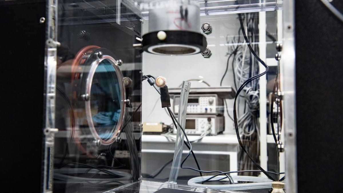
Biomedical Optical Imaging and Ultrasound laboratory (OPUS)
The University of Eastern Finland’s OPUS laboratory focuses on biomedical optical imaging and ultrasound research. The laboratory facilities are used by Optical Imaging team (Professor Tanja Tarvainen) in Inverse Problems group and by Biomedical Ultrasound group (Professor Kullervo Hynynen).
- Experimental diffuse optical tomography setup: Time-domain optical imaging of turbid samples. Tuneable wavelength nanosecond laser source and avalanche photodiode detector. Fibre-optic interface with 32 source fibres and 32 detector fibres.
- Experimental photoacoustic tomography setup: Optoacoustic imaging and characterization of biological and synthetic samples. Tuneable wavelength high-energy nanosecond laser source. Replaceable ultrasound transducer (bandwidth, geometry, size).
- Experimental photoacoustic microscope: Optical-resolution system using laser diode light source. Imaging of thin small samples.
- Hydrophone system: Computer-controlled 3D scanning station and fibre-optic hydrophone system for ultrasound pressure field measurements (Precision Acoustics).
- Radiation force balance: Ultrasound transducer acoustic power measurement.
- Schlieren imaging: System for measuring ultrasound beam patterns (Onda OptiSon).
- Laser Doppler vibrometer: Surface displacement and velocity measurements (Polytec).
- Ultrasound research imaging system: Verasonics Vantage 128™ system for high-speed ultrasound and photoacoustic imaging. 128 transmit channels and 128 receive channels. Linear array transducer. Raw RF data output.
- Optical detectors.
- Optical energy meters and sensors for pulsed and CW laser sources.
- Oscilloscopes and digitizers.
- RF signal generators and amplifiers.
- Ultrasound pulser-receivers.
- Ultrasound transducers.
- Ultrasound imaging phantom.
tanja.tarvainen@uef.fi
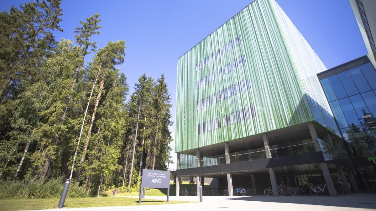
Computational Biophysics and Imaging Group
Tampere University’s Computational Biophysics and Imaging Group aims to deliver new knowledge and methods for future personalised medicine. Its facilities include 3D microstructure imaging instrumentation, bioimpedance, and cellular electrophysiology, complemented by open software tools developed in-house. CBIG is part of the Tampere Imaging Facility light microscopy core unit.
- X-ray microtomography: Zeiss Xradia MicroXCT-400
- Optical 3D-imaging microscopy: A Selective Plane Illumination Microscope (SPIM) and an in-house built Optical Projection Tomography (OPT) microscopes.
- Zeiss Axio Observer A1
- Sutter DG-4
- Olympus inverted microscope IX71
- Axopatch 200B amplifier
- Digidata 1440A Low-Noise data acquisition system
- Scientifica Patchstar micromanipulators
- Olympus inverted microscope IX73
- Axion Biosystems Maestro
- Multichannel Systems MEA2100
- Multichannel Systems MEA2100-lite
- Solartron impedance measurement device: System containing the Solartron 1260A Frequency Response Analyzer and 1294A Impedance Interface (Solartron Analytical, UK).
- HF2IS Impedance Spectroscope: System consists of the HF2IS impedance spectroscope and the HF2TA current amplifier (Zurich Instruments AG, Switzerland).
- Automatic objective neuronal spike detection for electrophysiological measurement
- CardioMDA: Used to analyse and evaluate drug response in cardiomyocyte cell population from field potential recordings.
- CorSE: Spectral entropy based neuronal network synchronisation analysis for extracellular electrophysiological data.
- ε-recurrence network analysis toolbox: The codes in the toolbox can be used to perform nonlinear time series analysis on single (or multi) channel data.
- Joint analysis of extracellular spike waveforms and neuronal network bursts
- SimpleCTC: Software that computes the contour tree connectivity (CTC) of 2D/3D images roviding, e.g., information on material porosity.
- Network-Wide Adaptive Burst Detection Depicts Neuronal Activity with Improved Accuracy: A modified version of the cumulative moving average (CMA) algorithm.
- Computational models of excitation-contraction coupling in human induced pluripotent stem cell-derived cardiomyocyte
jari.hyttinen@tuni.fi
Explore CBIG’s facilities on a virtual tour: https://www.thinglink.com/scene/1382993233621024769
Photo: Jenni Toivonen / Tampere University (cropped from the original)
DetectorLab
The Helsinki Institute of Physics Detector Laboratory at the University of Helsinki is a national permanent infrastructure specialized in the instrumentation of particle and nuclear physics. DetectorLab provides premises, equipment, and expertise for research projects on developing semiconductor, gas-filled and scintillator radiation detector technologies. Several position-sensitive semiconductor X-ray/gamma ray imaging devices, and relevant radioactive sources, are available.
- GeGI Germanium Gamma Ray Imaging Spectrometer: Portable gamma ray imager to characterize the radiological environment using multiple imaging modes. Automatic isotope detection, identification, localization, and source activity quantification make the GeGI useful in a variety of applications. The instrument provides list-mode data output, especially relevant for more fundamental imaging research.
- GDS-100 CZT-based detector: Contains two large cadmium-zinc-telluride (CZT) gamma ray detectors providing list-mode data output. The 3D position sensitivity makes this detector well-suited for gamma ray imaging.
- ADVACAM MiniPIX TPX3: Miniaturized particle tracking and X-ray/gamma imaging detector containing a 2 mm thick CdTe sensor on top of a Timepix3 readout (256 x 256 square pixels with a pitch of 55 μm).
Dr. Matti Kalliokoski
matti.kalliokoski@helsinki.fi
Photo: University of Helsinki / Linda Tammisto

Electrical tomography laboratory
The University of Eastern Finland’s Electrical tomography laboratory (ETL) is equipped with tomographic imaging devices, phantoms and accessories for imaging pipes, tanks, stirring vessels as well as cement-based materials and structures, ground, and human body. The tomographic devices are mainly based on electromagnetic interaction with the object, but also some other techniques, such as thermal stimulation are used.
- Process tomography section of the lab is equipped with tomography measurement systems and process simulators. The equipment can be used to examine various process applications such as multiphase flows, mixing and separation, and for the assessment of the condition of concrete and other solid structures.
- The flow pipe process simulator can be used to simulate, for example, liquid – liquid and liquid – gas mixing applications.
- The programmable logic system controls the main flow, chemical injection and gas injection.The logic systems consist of a programmable logic (Wago, type 750) and an industrial panel PC (IEI Technology). The logic system can be controlled via Ethernet or by local web user interface. The logic system also collects measurement data from conventional process measurement sensors.
- KIT4 Electrical impedance tomography measurement system: The latest EIT-device designed and constructed at the Department of Technical Physics. The system has 80 parallel voltage measurement channels and 16 parallel current injection channels. The maximum frame rate is 100 frames/second using 16 measurement channels. More detailed information about the KIT4 –device can be found in here.
- KIT 3 EIT measurement system: Designed mainly for conductivity probe measurements. It has 64 serial measurement channels and 4 parallel injection channels.
- Thermal tomography measurement system
- CoreApus Spectroscopic EIT measurement system
- Electromagnetic flow tomography (EMFT) measurement system: Electromagnetic flow tomography is an emerging imaging modality for estimating velocity fields of conductive flowing fluids in pipes. The approach behind the EMFT is the creation of magnetic field into the pipe flow, causing an electric field inside the pipe due to the moving charges. This electric field, or potential distribution, can be measured on the inside boundary of the pipe using a set of electrodes.
- SPIFIN Spectral induced polarization measurement system
- Datain Data infusion system
- PTL300E Electrical capacitance tomography system
- SRS830 DSP lock-in amplifier
- Hioki 3532-50 LCR -HiTester

Kuopio Biomedical Imaging Unit
Kuopio Biomedical Imaging Unit (Kuopio-BIU) provides open access services for biological research. It provides excellent facilities for small animal in vivo imaging and ex vivo MRI microscopy in different areas of basic and applied research. The imaging unit is located at the A.I.Virtanen Institute, University of Eastern Finland, and is open to both academic research and companies.
- MRI: Our equipment offers access to most common pre-clinical imaging platforms (Bruker, MR Solutions, Agilent). Most contemporary imaging and spectroscopy pulse sequences are available or can be implemented on request
- PET: μPET imaging is done in collaboration with Kuopio University Hospital and with its radiochemistry laboratory and cyclotron. Through this collaboration PET imaging with tailor made radiopharmaceuticals can be attained.
- CT: CT scanner can be used separately or in connection with the other imaging modalities.
- Optical, ultrasound and photo acoustic imaging: While not part of BIU, these services are also available upon request.
- Multimodal imaging: We have stereotactic small animal holders that can be used in the 7 T magnet, PET and the CT device allowing multimodality (MRI/PET/CT) imaging in the same coordinates.
- EEG/brain simulation/fMRI and awake fMRI: fMRI studies in animal models provide invaluable information regarding normal and abnormal brain function. Small animal fMRI studies are typically under anesthesia in order to minimize the stress and movement of the animals. However, anesthesia heavily modulates brain function and neurovascular coupling, making it probably the most serious confounding factor in preclinical fMRI. Kuopio-BIU has systematically studied the influence of anesthesia and can provide several different anesthesia protocols for different fMRI experiments. This expertise area is under active development.
- Metabolic MRI: Hyperpolarised metabolic MRI experiments can be combined with modern 1H imaging techniques to yield a comprehensive picture of the tissue status and gain a better understanding of potential biomarkers for metabolic diseases in brain and in other organs such as heart. This expertise area is under active development.
- Microstructural MRI: This is a multidisciplinary work in the application and combination of multiscale imaging modalities. Using the 11.7T MRI scanner the overall aim is to assess complex patterns of pathological tissue alterations in the brain using advanced magnetic resonance (MRI) techniques, and modern histopathological methods. This expertise area is under active development.
- PET/MRI & Kuopio Preclinical Molecular Imaging Platform: The platform combines the Kuopio University Hospital’s (KUH) radiotracer production with Kuopio-BIU and serves companies in the drug development cluster, clinical research groups at the University of Eastern Finland, and clinical research at KUH.
- Animal handling: The Kuopio BIU facility has complete anesthesia and physiological monitoring units suitable for in-bore monitoring of experimental animals.
- Data analysis: Most of our data analysis is done in Matlab and Python. Simple image analysis and calculation of e.g. relaxation time maps is performed using our home-built image analysis interface Aedes. For more complicated computing (Z-spectroscopy, diffusion tensor imaging…), separate analysis pipelines can be built. Functional and pharmacological MRI data is analyzed in FSL or SPM software and MRS data with LC model. Other analysis software for PET, MR and/or CT data include PMOD, Inveon Research Workplace and Carimas.
- Histology: Basic histological stainings.
- Behavioural studies: Basic behavioural studies on request.
Contact personnel (Kuopio-BIU homepage)
Photo: University of Eastern Finland / Törrönen Raija
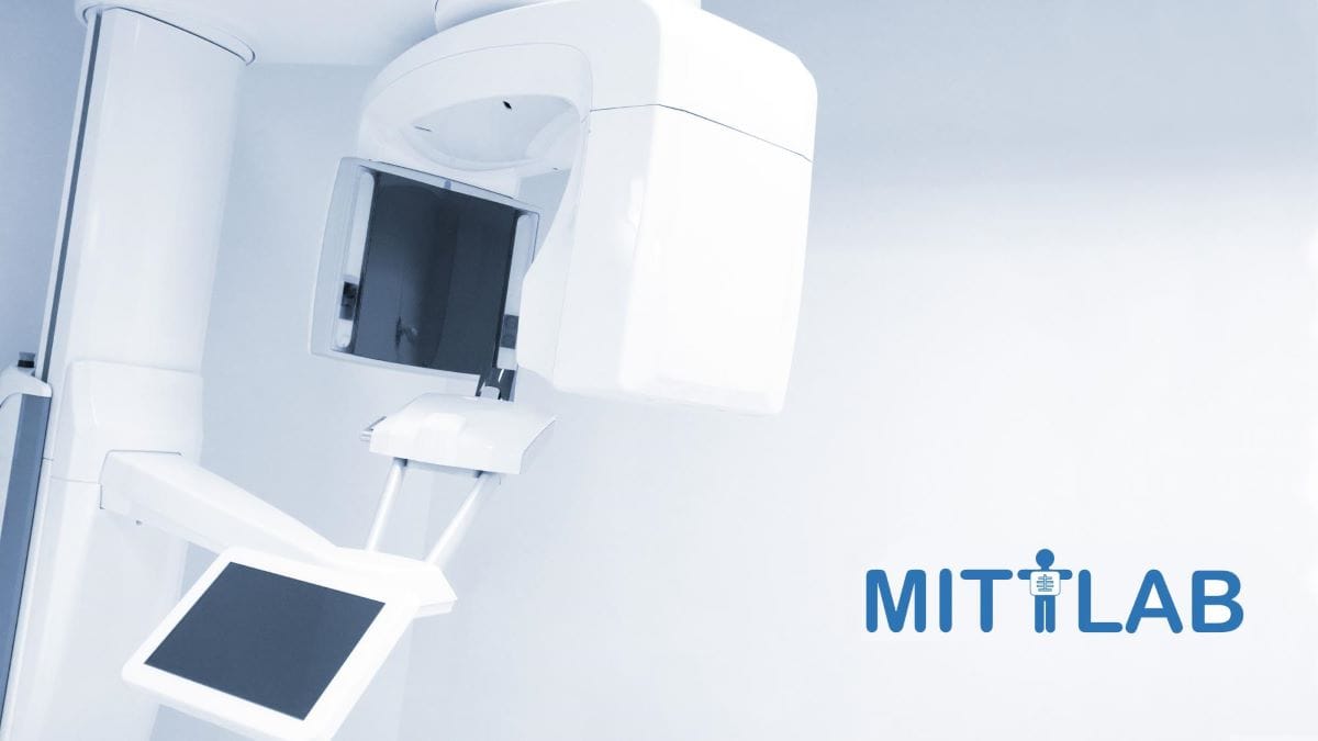
Medical Imaging Teaching and Test Laboratory (Mittlab)
Mittlab is a medical imaging teaching and test laboratory, that is dedicated to teaching, research, product development, innovation, and testing activities. Laboratory was established in a European regional development fund project in 2021 and is located at Oulu University Hospital. Part of OuluHealth Labs environment.
- Mammography: Siemens Mammomat Inspiration
- Dental CBCT: Planmeca Viso 7G
- Experimental CT-setup: Indented use is for tomographic imaging of biological samples and phantoms. X-ray tube, photon counting detector, and motorized rotator.
- Conventional CT: Toshiba Aquilion TSX-201A
- Conventional radiography: Fuji DR-XD 200X
- PIX-13: Leeds Test Objects’ PIX-13 phantom is used to check the image quality performance of digital radiography X-ray systems. These tests should be carried out at acceptance to establish a baseline, and on a routine basis, keeping an on-going record of the test results, which will indicate any deterioration in performance.
- AEC phantom: Leeds Test Objects′ TO AEC is an X-ray quality assurance phantom comprising a stack of PMMA plates which allow the user to check the consistency, repeatability and reproducibility of AEC system function.
- ACR phantom: The CIRS Model 086, ACR Digital Mammography (DM) Phantom was designed under the sponsorship of the American College of Radiology (ACR) to test the performance of FFDM systems. Objects within the phantom simulate calcifications, ducts and tumor masses. The phantom is designed to determine if your DM system can detect small structures that are important in the early detection of breast cancer.
- CTDI+ phantom: TO CTDI permits the user to measure the dose index for computed tomography systems. The test object consists of two PMMA cylinders, one cylinder represents the head (160mm diameter) and the other represents the body (320mm diameter). The length of each cylinder is 140mm. Each cylinder contains holes large enough to accept a radiation detector, used to take the dose measurements. There is a hole at the centre of the cylinders and one at each 90° interval, centered 10mm from the edge of the cylinder.
- TO MTF Edge Test Tool: An MTF Edge test device comprising an angulation wheel and a Tungsten edge.
- AI and Cu filters: 200x200mm Al and Cu filters, thicknesses 0.5, 1 and 2mm of Cu, and 1 and 2 mm of Al.
- Acryl plate: Leeds acryl plate 320*240 thickness 45mm.
- IBA patient equivalent: IBA patient equivalent 21mm (SN55), 21mm(SN54) and 25mm (SN380) Aluminum.
- CBCT phantoms and tools: Set of Planmecas’ phantoms and test tools to be used with our CBCT imaging device.
- Line pair patterns 38-010 & 39d-003: These test patterns are designed for quick quantitative assessments of the limiting spatial resolution of an X-ray system. The line pair patterns consist of a thin foil of Lead sandwiched between plastic plates. In the foil, linear slits are cut with a range of widths, these alternate with linear bars of the foil of equal width. One slit and one bar is referred to as a line pair, and the width of each group of lines is specified in terms of the number of line pairs per mm (LP/mm). The observer counts the number of groups in which he can resolve the slits from the bars and as such can determine the resolving capability of the X-ray system in terms of LP/mm.
- Piranha Premium Kit MULTI: Multipurpose all-in-one X-ray meter system for QA testing. Included: Dose probe for input dose to image intensifiers, scattered and leakage radiation. Light probe for measurements on monitors and for measurement of ambient light. CT Dose Profiler for CTDI measurements. MAS-1 probe for invasive mA and mAs.
- CTlab: CTlab is a virtual device interface of computed tomography (CT). Can be used for instance in teaching. Users are able to adjust many imaging parameters and see the effect on the image quality. Download: https://github.com/MIPT-Oulu.
- Virtual X-ray machine: Virtual X-ray machine is a virtual device interface of radiography. Can be used for instance in teaching. Users are able to adjust many imaging parameters and see the effect on the image quality. Download: https://github.com/MIPT-Oulu.
- MRiLab: MRiLab is a virtual device interface of magnetic resonance imaging (MRI). Can be used for instance in teaching. Users are able to adjust many imaging parameters and see the effect on the image quality. Download: https://github.com/MIPT-Oulu.
- X-ray scatter sim app: X-ray Scattering Simulator is a mobile application tool for demonstrating radiation scattering virtually. The demonstrated data is based on real life measurements in a hospital environment. The application is based on Augmented Reality technology (AR) that allows the user to add virtual 3D elements to the real-world environment through the camera of the mobile device. The objects in this application are the patient, X-ray machine and a color map depicting the radiation scattering. The purpose of this application is to demonstrate how the radiation generated during the X-ray imaging scatters to the environment. Link to the application: Google Play
Privacy Policy for X-ray Scatter Sim application
By using the app X-ray Scatter Sim you are consenting to our policies regarding the collection, use and disclosure of personal information set out in this privacy policy.
Collection of Personal Information: We do not collect, store, use or share any information, personal or otherwise.
Email: If you email the developer for support or other feedback, the emails with email addresses will be retained for quality assurance purposes. The email addresses will be used only to reply to the concerns or suggestions raised and will never be used for any marketing purpose.
Disclosure of Personal Information: We will not disclose your information to any third party except if you expressly consent or where required by law.
Contacting Us: If you have any questions regarding this privacy policy, you can email mittlab@lists.oulu.fi